Percutaneous Cholecystostomy
Updated: Dec 21, 2024
Sepsis and hypotension. What procedure is indicated? • Xray of the Week

Figure 1. What action should be taken for this patient with sepsis and hypotension?

Figure 2.
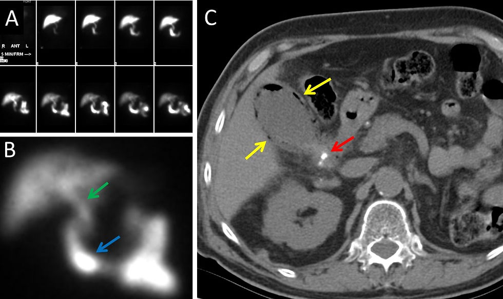
A. HIDA scan showing liver, common bile duct, small bowel
B. Magnified view HIDA scan showing common bile duct (green arrow) and duodenum (blue arrow). Note that tracer is not present in the gallbladder due to cystic duct obstruction.
C. Abdominal CT showing enlarged gallbladder with gas in the gallbladder wall (yellow arrow) and cholelithiasis (red arrow) indicating emphysematous cholecystitis.

Figure 3. Percutaneous Cholecystostomy
Abdominal CT showing percutaneous access needle (green arrow) entering the patient’s gallbladder via a transhepatic approach. The final CT image shows the drainage catheter (yellow arrow) correctly placed in the gallbladder with the tip coiled in the gallbladder fundus (red arrow).
Discussion:
Percutaneous cholecystostomy (PCS) is an image-guided, minimally invasive catheterization of the gallbladder [1]. It is indicated for gallbladder drainage in acute cholecystitis (including calculous, acalculous, gangrenous, and emphysematous varieties) or gallbladder perforation among high-risk patients with contraindications to surgical intervention. It can also be employed to allow percutaneous removal of biliary stones, or catheterization of the biliary tree to resolve obstruction [1,2]. Among patients treated for acute cholecystitis, PCS is commonly followed by cholecystectomy if the patient’s condition can be optimized, as its use as a definitive therapy in this population has not found consistent support [3].
PCS may be guided by US, CT, and/or fluoroscopy, and involves transhepatic or transperitoneal insertion of an access needle followed by gallbladder catheterization with either the Seldinger technique or a trocar system [1,4,5]. Figures 1 and 2 are imaging studies on a patient with sepsis and hypotension due to emphysematous cholecystitis who was too unstable to undergo surgery. The patient underwent a percutaneous cholecystostomy using the Seldinger technique and CT guidance, as illustrated in Figure 3. After stabilization and maturation of the tract, the patient had a surgical cholecystectomy and fully recovered.
Though ultrasound is typically the favored imaging modality for needle insertion due to mobility, lack of ionizing radiation, and continuous visualization, CT may be necessary if the gallbladder lumen cannot be observed sonographically (e.g. cholecystitis, wall thickening; 1,5). Access needles can be clearly visualized on either US or CT. Following US-guided needle insertion, fluoroscopy is frequently used to aid catheter placement [1,5].
Choice of transhepatic or transperitoneal approaches should be employed according to patient anatomy and operator discretion, since there has been no demonstrated difference in outcome or complications [6]. The former method involves traversing the liver with intent to puncture the bare area of the gallbladder, while the latter simply accesses the gallbladder via the peritoneal cavity [1,6]. Rationales favoring the transhepatic approach include increased catheter stability, quicker fistula tract maturation, and a theoretically decreased risk of bile leakage [1,6]. It is also preferred in cases of ascites or bowel interposition [1]. A transperitoneal route is favored in patients with coagulopathy or diffuse liver disease [1]. Major complications of PCS include hemorrhage, pneumothorax, biliary leak, and peritonitis, with the transhepatic approach having increased risk of pleural or hepatic damage [1,2,4].
References:
Ginat D and Saad W. Cholecystostomy and Transcholecystic Biliary Access. Tech Vasc Interv Radiol. 2008;11(1):2-13. DOI: 10.1053/j.tvir.2008.05.002
Hatzidakis A, Venetucci P, Krokidis M, Iaccarino V. Percutaneous biliary interventions through the gallbladder and cystic duct: What radiologists need to know. Clin Radiol. 2014;69(12):1304-1311. DOI: 10.1016/j.crad.2014.07.016
Gurusamy KS, Rossi M, Davidson BR. Percutaneous cholecystostomy for high risk surgical patients with acute calculous cholecystitis. Cochrane Database Syst Rev. 2013;(8): CD007088. DOI: 10.1002/14651858.CD007088.pub2
Little Mw. Percutaneous cholecystostomy: The radiologist’s role in treating acute cholecystitis. Clin Radiol. 2013;68(7): 654-660. DOI: 10.1016/j.crad.2013.01.017
Venara A, Carretier V, Lebigot J, E Lermite. Technique and indications of percutaneous cholecystostomy in the management of acute cholecystitis in 2014. J Visc Surg. 2014;151(6):435-439. DOI: 10.1016/j.jviscsurg.2014.06.003
Beland MD, Patel L, Ahn SH, Grand DJ. Image-Guided Cholecystostomy Tube Placement: Short- and Long-Term Outcomes of Transhepatic Versus Transperitoneal Placement. AJR Am J Roentgenol. 2019;212: 201-204. DOI: 10.2214/AJR.18.19669

Ian Rumball is a medical student and aspiring radiologist at the Zucker School of Medicine at Hofstra/Northwell in Hempstead, NY. He serves as chair for his school’s radiology interest group. Prior to medical school, he attended the University of Wisconsin - Madison and graduated with degrees in biology, history, global health, and African studies. As an undergraduate, he did research in the fields of oncology, hematology, and neuroendocrinology. He also published work in undergraduate journals of creative writing, history, and physiology. In his free time, Ian enjoys playing guitar, hiking his local state parks, and watching classic films.
Follow Ian Rumball on Twitter @RumballIan
UPDATE: Dr. Rumball will be a radiology resident at Medical College of Wisconsin in July 2024, after his Transitional Year at Gundersen Health System in La Crosse, Wisconsin.

Kevin M. Rice, MD is the president of Global Radiology CME and is a radiologist with Cape Radiology Group. He has held several leadership positions including Board Member and Chief of Staff at Valley Presbyterian Hospital in Los Angeles, California. Dr. Rice has made several media appearances as part of his ongoing commitment to public education. Dr. Rice's passion for state of the art radiology and teaching includes acting as a guest lecturer at UCLA. In 2015, Dr. Rice and Natalie Rice founded Global Radiology CME to provide innovative radiology education at exciting international destinations, with the world's foremost authorities in their field. In 2016, Dr. Rice was nominated and became a semifinalist for a "Minnie" Award for the Most Effective Radiology Educator. He was once again a semifinalist for a "Minnie" for 2021's Most Effective Radiology Educator by AuntMinnie.com. He has continued to teach by mentoring medical students interested in radiology. Everyone who he has mentored has been accepted into top programs across the country including Harvard, UC San Diego, Northwestern, Vanderbilt, and Thomas Jefferson.
Follow Dr. Rice on Twitter @KevinRiceMD


